
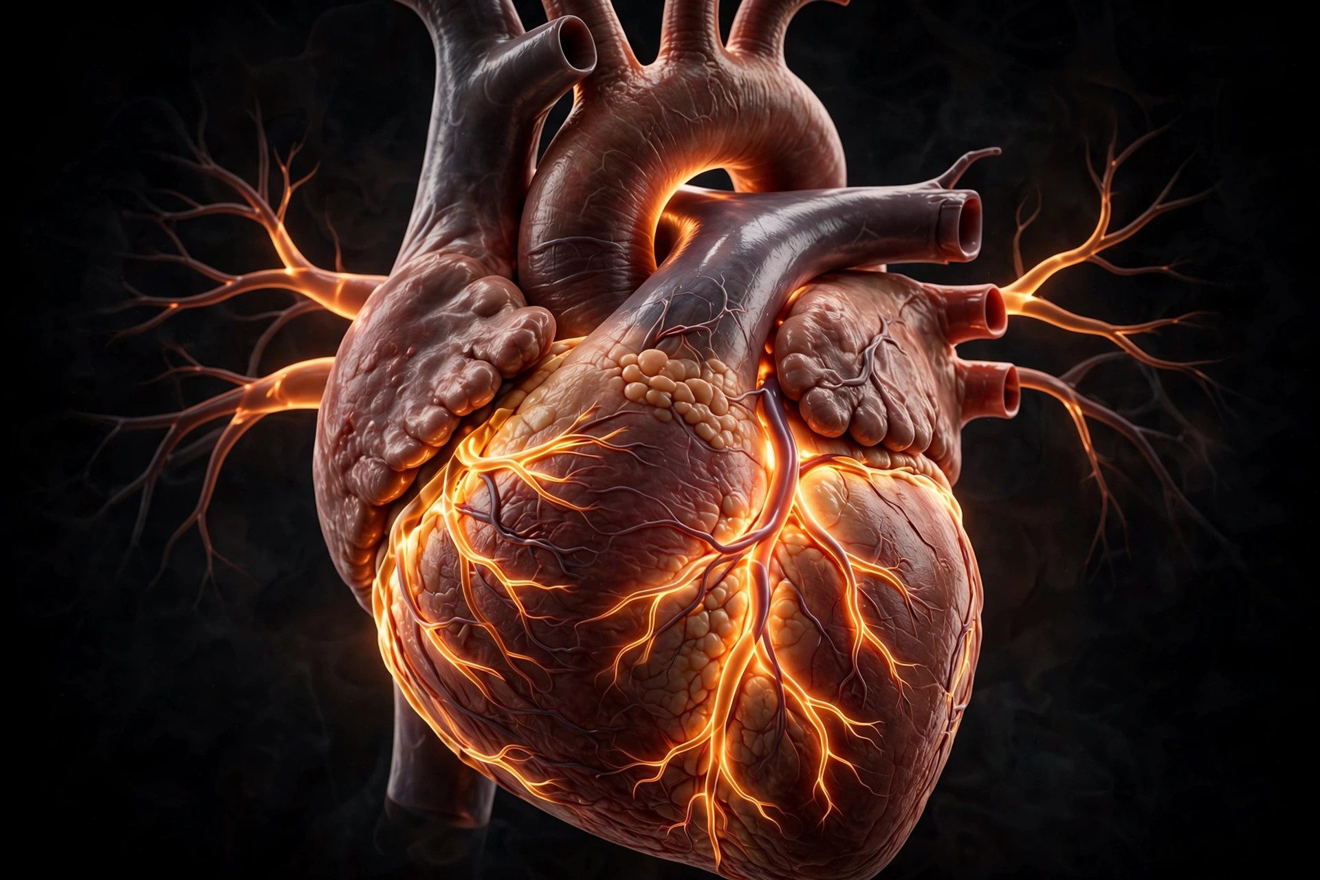
Your heart is a muscular organ roughly the size of your fist, sitting slightly left of center in your chest. Its job is simple to describe and astonishing to sustain: pump blood — carrying oxygen and nutrients — to every tissue in your body, and carry waste products away, continuously, for your entire life. It beats around 100,000 times a day and moves roughly 7,500 litres of blood daily, and it never gets a day off.
This guide explains how the heart works: its anatomy, the four chambers, the one-way valves, the built-in electrical system that sets the rhythm, and the cardiac cycle that repeats with every beat. You'll also learn what heart rate and blood pressure numbers mean, how the heart differs from every other muscle, and what keeps it healthy.
The heart's anatomy: built for a lifetime of work
The heart sits in the middle of the chest in a protective sac called the pericardium, which holds it in place and lets it beat without friction against surrounding structures. The heart wall itself has three layers: the thin outer epicardium, the thick muscular myocardium that does the pumping, and the smooth inner endocardium that lines the chambers and valves.
The myocardium is thickest around the left ventricle, because that chamber does the hardest job — pushing blood to the entire body. An adult heart weighs roughly 250–350 grams, about the same as a large apple, yet it generates enough force each day to lift a small car a short distance, if all its energy were concentrated in one burst.
The four chambers: a double pump
The heart has four chambers. The two upper chambers, the atria, receive incoming blood; the two lower chambers, the ventricles, pump it out with force. The right side handles oxygen-poor blood heading to the lungs; the left side handles oxygen-rich blood heading to the body. A muscular wall called the septum keeps the two sides completely separate — this "double pump" design is what makes warm-blooded, high-energy animals like us possible.
Follow the blood: two loops in one organ
Blood travels through the heart in a precise sequence, completing two connected loops:
The pulmonary loop (heart → lungs → heart)
Oxygen-poor blood returning from the body enters the right atrium through two large veins, the venae cavae. It flows down into the right ventricle, which pumps it through the pulmonary valve into the pulmonary arteries — the only arteries in the body that carry oxygen-poor blood — toward the lungs. There, carbon dioxide is released and fresh oxygen is picked up.
The systemic loop (heart → body → heart)
Oxygen-rich blood returns from the lungs through the pulmonary veins into the left atrium, drops into the left ventricle, and is then forced through the aortic valve into the aorta — the largest artery in the body. From the aorta, blood branches into ever-smaller vessels that reach every cell, delivering oxygen and nutrients and collecting waste before the journey begins again.
Valves: the heart's one-way doors
Four valves act as one-way doors between chambers and vessels, opening and closing with each beat to prevent backflow. The tricuspid and mitral valves sit between the atria and ventricles; the pulmonary and aortic valves guard the exits to the lungs and body. The familiar "lub-dub" sound of the heartbeat is largely the sound of these valves snapping shut — "lub" when the atrioventricular valves close, "dub" when the semilunar valves close.
The electrical system: your built-in pacemaker
Each heartbeat is triggered by a tiny electrical signal that begins in the sinoatrial (SA) node — the heart's natural pacemaker — in the right atrium. The signal spreads across the atria, making them contract and push blood into the ventricles. It then pauses briefly at the atrioventricular (AV) node, giving the ventricles time to fill, before racing down specialized fibres (the bundle of His and Purkinje fibres) that make the ventricles contract from the bottom up — the most efficient direction for pumping.
This coordinated sequence is what an electrocardiogram (ECG) records. Doctors read the ECG's waves to check whether the rhythm is normal or whether the electrical system is misfiring, as in an arrhythmia. When the natural pacemaker fails, an artificial pacemaker — a small implanted device — can take over the signalling job.
The cardiac cycle: systole and diastole
One full beat has two phases. During systole, the heart muscle contracts and pushes blood out of the chambers. During diastole, it relaxes and the chambers refill. At rest the cycle repeats roughly once per second; it speeds up with exercise, excitement or stress and slows during rest and sleep. Interestingly, the heart muscle itself receives its blood supply during diastole, through the coronary arteries — which is why a racing heart under strain can struggle to feed itself.
How the heart differs from every other muscle
Heart muscle (cardiac muscle) is unlike skeletal muscle in several ways. Its cells are connected by special junctions that let electrical signals pass directly from cell to cell, so the whole chamber contracts as one unit. It has an enormous density of mitochondria — the cell's energy factories — and a rich blood supply, because it can never stop to rest. Skeletal muscles tire; the heart is built to contract rhythmically for a century without fatigue.
Heart rate and blood pressure: what the numbers mean
A typical resting heart rate for adults is 60–100 beats per minute, with trained athletes often resting below 60. Blood pressure is written as two numbers, such as 120/80: the upper (systolic) number is the pressure when the heart contracts, and the lower (diastolic) number is the pressure when it relaxes. Both numbers matter, and both tend to rise temporarily with activity, stress, caffeine or excitement — what counts for health is the long-term pattern, not a single reading.
Common heart conditions, briefly
The most common heart problems involve the plumbing or the electrics. Coronary artery disease happens when the coronary arteries narrow, reducing blood flow to the heart muscle itself. Arrhythmias are rhythm disturbances — the heart beating too fast, too slow or irregularly. Heart failure means the pump has weakened and can't move blood as efficiently as the body needs. These are medical conditions that need professional diagnosis; this article is educational, not a diagnostic tool.
Keeping the pump healthy
Health authorities consistently emphasize the same foundations: regular physical activity, a balanced diet rich in vegetables, fruits and whole grains, not smoking, maintaining a healthy weight, and managing blood pressure and cholesterol. Aerobic exercise strengthens the heart muscle itself, while good sleep gives the cardiovascular system its nightly recovery window. Warning signs such as chest pain or pressure, unusual breathlessness, or fainting deserve prompt medical attention — never ignore them.
Fascinating facts about the heart
- The heart starts beating around three to four weeks after conception — before most people know they're pregnant.
- A woman's heart typically beats slightly faster than a man's at rest.
- The heart can keep beating even outside the body for a short time, because it generates its own electrical impulses.
- Laughter is genuinely good for the heart: it temporarily improves blood vessel function.
- The "break" in a broken heart isn't just poetic — extreme emotional stress can temporarily weaken the heart muscle, a real condition called stress cardiomyopathy.
That last point is why stress management counts as heart care: practices such as mindfulness — short, structured exercises in paying attention — are a popular way to keep everyday stress in check.
This article is for general information only and is not medical advice. If you have concerns about your heart, speak to a qualified healthcare professional.